Ligaments Of The Knee Anterior / Exam Series Guide To The Knee Exam Canadiem / The anterior cruciate ligament sits deep in the middle of the knee joint.
Ligaments Of The Knee Anterior / Exam Series Guide To The Knee Exam Canadiem / The anterior cruciate ligament sits deep in the middle of the knee joint.. Perhaps the earliest account of the all was written by french surgeon paul segond in 1879, in which he described a ligamentous structure between the lateral femur and tibia. From wikipedia, the free encyclopedia the anterolateral ligament (all) is a ligament on the lateral aspect of the human knee, anterior to the fibular collateral ligament. It runs upward, backward, and laterally and is connected to theposterior part of the medial surface of the lateral condyle of femur. Two of these ligaments are in the center of the joint, and they cross each other. The acl is a tough band of tissue joining the thigh bone to the shin bone at the knee joint.
Ligament is what the medicine world calls the tough bands of tissue that connect bones or hold organs in place. There are four major ligaments that surround the knee joint. Typically, your knee will immediately start to swell up. The anterior cruciate ligament (acl) and the other being the posterior cruciate ligament (pcl). Bones, cartilage, ligaments, and tendons.

The kneecap slides along a groove in the femur as the knee bends.
The anterior cruciate ligament (acl) is a band of dense connective tissue which courses from the femur to the tibia. This topic review will discuss the presentation, evaluation, and management of acl injuries. It is held in place by a ligament at the bottom and a. The anterior cruciate ligament runs diagonally in the middle of the knee. Both (acl & pcl) are present inside your knee joint. Diagnostic evaluation of the knee. The anterior cruciate ligament (acl) is essential to keeping the knee stable and balanced. The anterior cruciate ligament sits deep in the middle of the knee joint. Cross your long fingers over the index finger and superimpose this hand over your ipsilateral knee. Also remember the mnemonic lamp which means lateral acl. Knee ligaments are comprised of soft tissue that connects bone to bone. It runs upward, backward, and laterally and is connected to theposterior part of the medial surface of the lateral condyle of femur. The anterior cruciate ligament (acl) is an important stabilizing ligament of the knee that is frequently injured by athletes and trauma victims.
Ligament is what the medicine world calls the tough bands of tissue that connect bones or hold organs in place. Ligaments are strong bands of tissue that connect one bone to another. The anterolateral ligament (all) of the knee has gained attention recently for its potential role in rotational stability of the knee, especially in association with anterior cruciate ligament (acl) injuries. Injuries to the anterior cruciate ligament ( acl ), posterior cruciate ligament ( pcl ), medial collateral ligament ( mcl ), and lateral collateral ligament ( lcl ) result in knee pain and instability. The extracapsular ligaments include the patellar, medial collateral, and oblique and arcuate popliteal.
Anterior cruciate ligament (acl) injury your acl connects the inside of the top of your tibia (shinbone) to the outside bottom of your femur (thighbone) in the front of the knee.
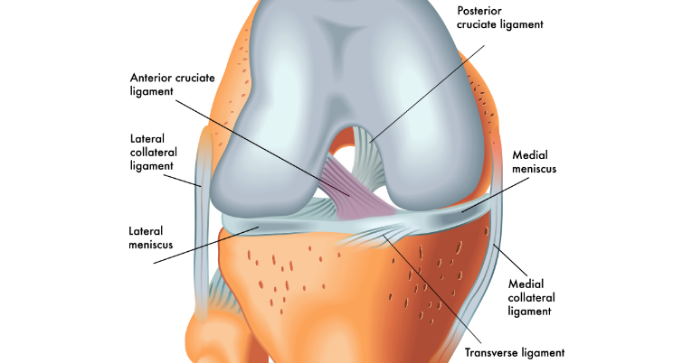
These knee ligaments help stabilize and strengthen the largest joint in the body by limiting its range of motion. The anterior cruciate ligament (acl) is an important stabilizing ligament of the knee that is frequently injured by athletes and trauma victims. Ligaments join the knee bones and provide stability to the knee: The anterolateral ligament (all) of the knee has gained attention recently for its potential role in rotational stability of the knee, especially in association with anterior cruciate ligament (acl) injuries. This ligament is almost twice as strong and has better blood supply than the anterior cruciate ligament. The knee is stabilized by a pair of cruciate ligaments. Knee ligaments are comprised of soft tissue that connects bone to bone. The anterior cruciate ligament (acl) is a band of dense connective tissue which courses from the femur to the tibia. Knee ligament injuries are often the result of rotational movement of the knee joint (e.g., cutting and pivoting movements in sports). Both (acl & pcl) are present inside your knee joint. The transverse ligament of the knee, also called the transverse intermeniscal ligament attaches transversely across the anterior aspects of the convex margins of the medial and lateral menisci. The anterior cruciate ligament prevents the femur from. The four key ligaments of the knee are:
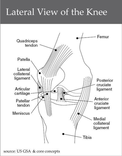
This will help us to remember the orientation of the anterior cruciate ligament (acl) and posterior cruciate ligament (pcl) of knee. These are called the cruciate ligaments and consist of the anterior cruciate ligament and the posterior cruciate ligament. There are two cruciate ligaments present in the knee joint: Two of these ligaments are in the center of the joint, and they cross each other. The anterior cruciate ligament (acl) is one of the 4 major ligaments of the knee.
When the anterior drawer test is done, if an audible snap or palpable jerk (finochietto jumping sign) occurs when the tibia is pulled forward, and the tibia moves forward excessively, a meniscal lesion is likely in addition to the torn anterior cruciate ligament.
The anterior cruciate ligament (acl) and the other being the posterior cruciate ligament (pcl). Ligament is what the medicine world calls the tough bands of tissue that connect bones or hold organs in place. This will help us to remember the orientation of the anterior cruciate ligament (acl) and posterior cruciate ligament (pcl) of knee. These knee ligaments help stabilize and strengthen the largest joint in the body by limiting its range of motion. The four main ligaments in the knee connect the femur (thighbone) to the tibia (shin bone), and include the following: Diagnostic evaluation of the knee. It attaches to the front of the tibia and the back of the femur. Your knee can buckle and cause pain. There are four major ligaments that surround the knee joint. Acl crosses over the pcl to form an 'x' shape inside your knee, signifying acl lies in front and pcl lies backward. Gross anatomy the acl arises from the anteromedial aspect of the intercondylar area on the tibial plateau and passes upwards and backwards to. If you tear the anterior cruciate ligament (acl) in your knee, you may need to have reconstructive surgery. The acl is a tough band of tissue joining the thigh bone to the shin bone at the knee joint.


Komentar
Posting Komentar